
Breast cancer develops when there is uncontrolled growth of the structure that forms the milk ducts and glands in the breast. Breast cancer is estrogen-dependent and is an endocrine tumor.
It is the most common cancer type among women, and in many countries, including Turkey, it ranks as a leading cause of death. However, when treated appropriately, while its incidence is high, its fatality rate is relatively lower.
In Turkey, breast cancer is among the top 10 most frequently occurring cancer types in women. The breast is made up of lobules that produce milk and ducts that transport the milk to the nipple. Breast cancer develops when there is uncontrolled growth of these two structures.
Cancer that originates in the lobules is called lobular cancer, while cancer that develops in the milk ducts is referred to as ductal cancer. Ductal cancer is the most common type and makes up the majority of breast cancers.
Breast cancer is the most common cancer in women. Worldwide, on average, one in every 8 women is diagnosed with breast cancer. The incidence of breast cancer in Turkey is similar to the global average.
The causes and risk factors of breast cancer can be categorized into controllable and uncontrollable factors.
Uncontrollable risk factors:
Please note that these are uncontrollable risk factors for breast cancer, and it's essential to be aware of them for early detection and prevention.
Controllable Risk Factors:
Maintaining a healthy lifestyle, including a balanced diet, limited alcohol consumption, and regular physical activity, can help reduce the controllable risk factors associated with breast cancer. Regular medical check-ups and screenings are also crucial for early detection and prevention.
1. Breast Lump: A significant finding in many breast cancer patients is the presence of a lump in the breast. Most lumps are painless, but they can also be associated with pain. They are most commonly found in the upper outer quadrant of the breast because the majority of breast tissue is located there. These lumps often have poorly defined borders and may feel rough or irregular. While the majority of lumps detected in the breast are benign, every breast lump should be considered potentially cancerous until proven otherwise, and necessary investigations should be conducted.
2. Nipple Discharge: Spontaneous, unilateral, and bloody nipple discharge may be associated with cancer in approximately 10% of cases, and any nipple discharge should be taken seriously and evaluated. A cytological examination should be performed on nipple discharge for pathological evaluation.
3. Pain: Pain in breast cancer is more commonly observed in the later stages of the disease. Pain associated with cancer occurring without any other clinical symptoms is very rare. It is typically seen in benign breast conditions. Breast pain is an important symptom and is one of the complaints that should promptly lead the patient to seek medical attention.
4. Nipple Retraction: It is mostly observed in cancers located near the nipple. However, this appearance can also be structural and non-pathological. New, unilateral nipple retraction should always be evaluated as a potential sign of cancer.
5. Edema of the Breast Skin: Thickening of the breast skin and a "peau d'orange" (orange peel) appearance can result from cancer cells blocking lymphatic vessels. This is a sign of advanced breast cancer and is usually observed in later stages.
6. Eczema/Rash on the Skin Around the Nipple: Lesions caused by irritation usually improve within 1-2 weeks with appropriate treatment, while lesions associated with cancer do not improve and require evaluation.
7. Lump in the Armpit: This occurs when the tumor spreads through the lymphatic vessels to the lymph nodes in the armpit. It should be investigated without delay.
Recognizing and addressing these symptoms is crucial for early detection and prompt medical evaluation.
The incidence of breast cancer is steadily increasing; however, the chances of early detection are also high, and when diagnosed early, patients have a significant chance of overcoming this disease. One of the major factors in early diagnosis is self-breast examination. When diagnosed early, the treatment can usually be administered effectively.
Women can typically detect a mass that has reached a size of 1 cm. In addition to self-breast examination, medical examinations, and imaging techniques for the breast play a significant role in early diagnosis. Imaging methods in breast cancer are important for detecting smaller, non-palpable, and unnoticed cancers
Approximately 1 gram of breast cancer tissue takes an average of 8 years to develop. In other words, it takes about 5 years for a 1 cm breast cancer to form. If breast cancer is left untreated, it can lead to distant organ metastases and result in death within 5 years.
Early-stage breast cancer is treatable, but in advanced stages with distant organ metastasis, treatment becomes challenging. Breast cancer treatment requires a multidisciplinary approach. In breast cancer, the goal of early diagnosis is to detect the disease clinically before it presents symptoms, shortly after its biological onset.
Early diagnosis is essential for the successful management and treatment of breast cancer. Regular screenings, self-examinations, and prompt medical attention for any concerning symptoms are vital for early detection and better outcomes.
Breast cancer can be detected through various imaging methods, including:
1. Mammography
After a clinical examination, additional imaging methods may be necessary, and mammography is often the primary choice for breast imaging. Mammography is the most effective method for the early detection of breast cancer.
Mammography is a technique that uses a low-dose X-ray to capture images of the breast. The goal is to detect breast cancer as early as possible. This imaging method involves compressing the breast between two layers and taking images from top to bottom and from side to side. It is known to be a painful procedure for some women.
In the hands of experienced professionals, mammography should not cause unbearable pain, and the quality of the mammogram is essential. Every woman, regardless of whether they have symptoms or not, should have a mammogram taken annually starting at the age of 40. This is crucial for early diagnosis and is an indispensable imaging method for patient follow-ups.
2. Breast Ultrasound
Ultrasound is an imaging method that utilizes sound waves to create images. The images are formed by the reflection of sound waves on a screen and are used to assess structures within the breast. Ultrasound is particularly useful for distinguishing between solid and cystic formations within the breast.
One of the significant advantages of breast ultrasound is that it does not involve radiation, making it a safe and comfortable procedure that can be performed as often as needed. It is also safe for pregnant and breastfeeding women. Breast ultrasound is commonly recommended for individuals with dense breast tissue detected in mammography. It is one of the examination methods that require the most experience.
To ensure that findings are not overlooked or misinterpreted, experience and expertise are crucial. The evaluation of the patient by experienced breast radiologists is also highly important in the process.
3. Magnetic Resonance Imaging (MRI)
MRI, also known as magnetic resonance imaging or simply MRI, is used for the identification of mammary abnormalities. It is typically employed in high-risk individuals and those who have recently been diagnosed with breast cancer. It may be preferred for individuals with dense breast tissue and those for whom mammographic evaluation is not suitable. If a lesion is visible only on MRI, a biopsy should be performed without hesitation. MRI can also be used to evaluate the recurrence of breast cancer in cases where treatment has already been administered.
4. Positron Emission Tomography (PET)
PET is a relatively new cancer diagnostic method, and research in this field is still ongoing. In this procedure, a radioactive substance is administered to the patient. Cancer cells are known to be rapidly dividing cells, and they tend to accumulate the administered radioactive material more quickly than other cells. This method is an imaging technique that identifies the potential location of cancer cells.
PET imaging is used to determine the spread (metastasis) of cancer, detect whether cancer is present in lymph nodes, and assess the status of cancer after treatments such as chemotherapy and radiotherapy, which are part of the treatment plan. PET is not typically preferred as a replacement for mammography or ultrasound. It is not used for breast cancer screening.
5. Tomosynthesis
Tomosynthesis is a relatively recent imaging technique that allows for the evaluation of breast tissue in three dimensions. With this method, numerous images are captured from different angles, and a computer system is used to assess the breast tissue in millimeter-thick slices, similar to a tomographic scan.
The treatment of breast cancer requires a multidisciplinary approach. While surgical treatment forms the initial stage of breast cancer treatment, other treatments may take priority based on the size, spread, and type of the tumor. Surgery and radiotherapy provide local control, while chemotherapy and hormone therapy offer systemic control. Psychological counseling is an integral part of breast cancer management.
The city of Ankara, like many other major cities, offers comprehensive breast cancer treatment options. Ankara is home to numerous medical facilities and specialized oncology centers that provide a wide range of services for breast cancer patients. These services include surgery, radiation therapy, chemotherapy, hormone therapy, and psychosocial support.
Patients diagnosed with breast cancer in Ankara should consult with their healthcare providers to determine the most suitable treatment plan tailored to their specific condition. Treatment decisions are typically made in consultation with a team of medical professionals, including surgeons, oncologists, radiologists, and psychologists, to ensure the best possible care for the patient.
When deciding on the treatment for breast cancer, all treatment options are discussed with the patient. The success of breast cancer treatment is closely related to early detection of the disease. Taking a thorough medical history, conducting a physical examination, and performing the necessary imaging studies are essential.
After these steps, a diagnosis is established through biopsy in eligible cases, and staging is carried out. Staging is the process of determining the extent of the disease at the time of diagnosis. It helps guide the treatment plan and decisions regarding the course of the disease.
Mastectomy is the surgical removal of the entire breast tissue. It is performed when breast-conserving surgery is not an appropriate option. If the patient has lymph node involvement in the axillary region, the removal of the entire breast along with axillary lymph nodes is carried out, and this is known as a modified radical mastectomy.
This is a traditional surgical method that provides good local control, and the risk of tumor recurrence is low. However, it is not a method preferred by many patients for aesthetic reasons. Although this surgical approach may initially seem acceptable to patients, it can lead to significant aesthetic concerns for them after breast cancer treatment is completed.
The primary goal of breast-conserving surgery is to treat breast cancer while achieving a good cosmetic outcome after surgery. It is considered the standard treatment for early-stage breast cancers today. Early detection methods, increased patient awareness, and the ability to catch the disease in its early stages have made breast-conserving surgery possible. Following this surgery, radiotherapy is typically planned for the patient.
Complete removal of the breast in patients can lead to psychological trauma, including depression, emotional disturbances, loss of sexual desire, decreased body image, loss of femininity, and concerns about the disease's recurrence. Breast-conserving surgery is a procedure in which the breast is preserved, and only the cancerous portion is removed, provided the patient's acceptance, the suitability of the breast, and the appropriateness of the tumor. During the operation, pathology is examined to ensure that surgical margins are negative.
To perform breast-conserving surgery, the patient must be informed about all surgical procedures, and the patient must be willing to accept all necessary treatments. The remaining breast after breast-conserving surgery should be aesthetically satisfying, and the patient should have access to a center where radiotherapy can be administered because radiotherapy is always required after breast-conserving surgery.
The lymph drainage in the breast primarily flows towards the axillary (armpit) lymph nodes. Therefore, the status of the axillary lymph nodes in breast cancer is crucial for staging, treatment, and follow-up. If the tumor has spread to the axillary lymph nodes, it necessitates the removal of these nodes. In the SLNB procedure, a blue dye (isosulfan blue or methylene blue) alone or in combination with a radioactive substance (ROLL) is injected into the area of the tumor or just beneath the nipple. After waiting for about 10-15 minutes, when the blue dye or radioactive substance reaches the axillary lymph nodes, the sentinel lymph node, the first lymph node in the axilla, is identified by the presence of the dye or radioactive material. There can be more than one sentinel lymph node, and during surgery, a pathology examination determines whether they contain tumor cells.
If tumor cells are not found, the procedure is concluded, thus preventing unnecessary surgery. If tumor cells are present, the axillary lymph nodes are cleared. The involvement of axillary lymph nodes is an important indicator of how cancer will progress. Patients who have had their axillary lymph nodes removed should take special care to protect the arm from swelling (lymphedema) that may occur. This includes careful hygiene, avoiding trauma to the arm, and taking precautions when drawing blood or measuring blood pressure from that arm.
To ensure that the axillary lymph nodes are properly sampled, an average of about 10 lymph nodes may need to be removed. These patients should be protected from arm trauma or infection. The incidence of lymphedema after SLNB is quite low. After the procedure, patients should be encouraged to perform physical exercises. In some cases, lymph fluid may accumulate in the armpit after axillary lymph node dissection. If it causes discomfort, it can be easily drained using an injection with ultrasound guidance. There's no need for drainage unless it bothers the patient.
Breast reconstruction surgery has been shown to have positive effects on the psychological well-being of patients. It allows for follow-up without any significant issues. It has no negative impact on the recurrence of the disease or the success of the treatment.
Breast reconstruction using only the patient's own tissue is called autologous reconstruction. It is mainly performed using tissue samples taken from the abdomen, buttocks, back, and inner thighs. In addition to this method, reconstruction with implants (silicone breast reconstruction) can also be performed. These can be permanent, fixed breast implants or temporary tissue expanders.
The most commonly used are permanent, fixed breast implants. These are silicone-filled implants with a textured outer surface. However, if the patient requires postoperative radiotherapy, this may not be the best option. In this procedure, if possible, the nipple is preserved, the breast skin is preserved, the entire breast is removed, and a silicone breast implant is placed under the muscle or under the skin to complete the procedure. This also helps prevent any psychological trauma that patients may experience after the surgery.
The goal of risk-reducing surgery is to reduce the risk of developing breast cancer in individuals at high risk for the disease by removing breast tissue before cancer develops. Advances in genetic testing methods and understanding of familial inheritance, as well as the identification of genetic mutations that lead to breast cancer, allow for a better estimation of a patient's likelihood of developing breast cancer. Such patients should undergo thorough genetic testing to make an informed decision.
Afterward, all the details should be discussed with the patient, and any uncertainties should be addressed. In risk-reducing surgery, after a bilateral subcutaneous mastectomy, reconstruction can be performed using implants (silicone),which not only significantly reduces the risk of breast cancer but also allows the patient to continue life with an aesthetically pleasing breast.
Radiation therapy is a treatment method that uses X-rays. It is performed with the aim of achieving local control of the tumor, much like surgery. It can be used on its own or in conjunction with chemotherapy, either before or after surgery. It is necessary in cases where breast-conserving surgery is performed, and it has been reported to reduce the risk of tumor recurrence. If the entire breast has been removed (mastectomy),or if the tumor is adhered to the chest wall or the breast skin, or if there are more than 4 lymph nodes under the arm involved, radiation therapy is recommended.
Radiation therapy should be administered within six months after the completion of surgical wound healing. If this period is extended, the chances of success decrease. If chemotherapy is needed during this period, it can either be delayed until after radiation therapy or a break in chemotherapy can be taken to administer radiation therapy, followed by a resumption of chemotherapy.
The goal of radiation therapy is to prevent the growth and proliferation of cells in the treated area. During radiation therapy, both cancer cells and normal cells are affected, but normal cells typically recover after treatment. If radiation therapy is administered to the armpit, there may be swelling (lymphedema) in the arm. In suitable centers, radiation therapy can also be administered during surgery.
Chemotherapy targets rapidly growing cells, including cancer cells, with the goal of halting their growth or destroying them. Chemotherapy can be administered using a single drug or, more commonly, a combination of multiple drugs. The aim here is to provide systemic treatment.
Chemotherapy is the primary treatment approach for advanced disease (in the presence of metastasis). It typically begins around 1 month after surgery, allowing time for the surgical wound to heal. A preferred chemotherapy regimen usually consists of an average of 4-6 cycles. There is an average of 2-3 weeks between each cycle, and blood tests are performed before each treatment because chemotherapy can have adverse effects on bone marrow and the immune system. The benefits of chemotherapy outweigh the potential risks, and any side effects that may occur are generally temporary.
After surgical treatment and chemotherapy, if the evaluation of tissue samples taken after surgery shows that the cancer is hormone-sensitive (responsive to estrogen and progesterone),hormone drugs are administered. This treatment is known as hormone therapy. The primary source of estrogen and progesterone hormones in the body is the ovaries.
Because breast cancer is hormone-sensitive, hormone therapy targeted at these hormones has been found to have a protective effect against breast cancer. Therefore, the most commonly used drug is Tamoxifen. In addition to Tamoxifen, Aromatase Inhibitors are also used. Tamoxifen is typically used for 5 years for preventive purposes, but it can cause symptoms similar to menopause.
The long-term use of Tamoxifen may also increase the risk of uterine cancer, so it is limited to 5 years of use. Aromatase Inhibitors given after menopause can have side effects such as bone loss and joint pain. Patients taking these medications should have their bone density measured, take calcium supplements, and receive physical therapy if necessary.
In breast cancer treatment, targeted therapies work through a different mechanism than traditional chemotherapy drugs. These drugs specifically target cancer cells and minimize damage to healthy cells, potentially resulting in fewer side effects.
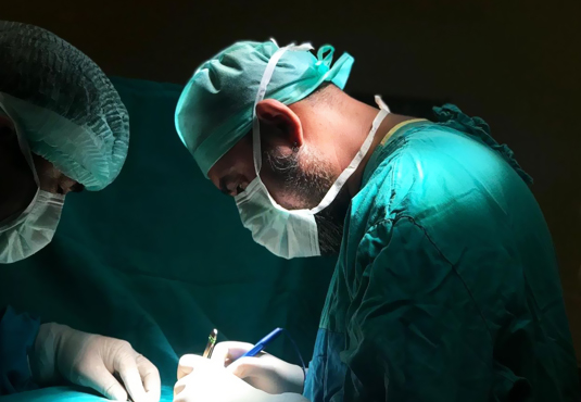
Breast cancer surgery is a surgical procedure performed on patients who have been diagnosed with breast cancer following the detection of a suspicious lesion in the breast and a subsequent biopsy. The specific type of surgery depends on factors such as the size of the cancer, the size of the lesion identified, the involvement of the axillary lymph nodes, and whether the cancer has spread to other organs. The surgery is performed as part of the treatment plan for breast cancer patients.
The choice of breast cancer surgery method is determined based on factors such as the size of the breast cancer, the involvement of axillary lymph nodes, the presence of metastasis in other organs, the patient's expectations, and their overall condition. The surgical methods for breast cancer include the following:
To perform breast-conserving surgery, the cancerous mass in the breast should be relatively small in proportion to the breast size. This allows for the removal of the cancerous mass along with the surrounding healthy tissue, leaving a cosmetically acceptable breast size. Additionally, breast-conserving surgery is typically followed by radiotherapy, so the patient should be informed about this radiotherapy treatment and have easy access to a radiotherapy center. Another crucial aspect is that the patient must consent to this treatment. When all these conditions are met, it is possible to perform surgery that leaves the breast in place while removing the tumor-containing mass along with the surrounding healthy tissue. This procedure is called breast-conserving surgery.
Is Breast-Conserving Surgery (Operation) Suitable for Everyone?
Breast-conserving surgery (operation) may not be suitable for every patient. After the patient has accepted breast-conserving surgery, the breast volume (size) and the size of the mass should be suitable for breast-conserving surgery. Additionally, because radiotherapy treatment will be administered afterward, the patient should have easy access to a facility where radiotherapy can be performed. When all these conditions are met, breast-conserving surgery can be performed on the patient.
Simple mastectomy is a surgical method for breast cancer in cases where there is rarely lymph node spread to the armpit and no distant organ metastasis. Simple mastectomy can involve the removal of the entire breast, including the nipple, areola, and breast skin, or it can be a skin-sparing simple mastectomy, which is preferred in cases where breast reconstruction with a breast prosthesis is planned. Skin-sparing simple mastectomy preserves the nipple, areola, and breast skin while removing the entire breast.
What Is a Mastectomy?
A mastectomy is a surgical procedure performed under general anesthesia to remove the entire breast, often including the nipple. If breast reconstruction with a breast prosthesis (implant) is planned, a skin-sparing mastectomy surgery can be performed to preserve the breast skin, with or without preserving the nipple.
Modified radical mastectomy is a surgical procedure performed on breast cancer patients with axillary lymph node involvement, in which the breast tissue is completely removed over the chest muscle without preserving the nipple, areola, and breast skin. This surgery also includes axillary dissection, which is the removal of the lymph nodes in the armpit. The combination of these two procedures is referred to as modified radical mastectomy.
Mastectomy + oncoplastic surgery is a surgical procedure for breast cancer patients in which the entire breast is removed, and the resulting void is filled with healthy tissue and muscle from another part of the patient's body. This approach is used to reconstruct the breast after a mastectomy.
Mastectomy + reconstruction with implants is a surgical procedure used for breast cancer patients or those at high risk of breast cancer due to genetic factors. It involves removing the entire breast tissue while preserving the nipple, areola, and breast skin (referred to as skin-sparing mastectomy) and then reconstructing the breast by placing a silicone breast implant for a good aesthetic outcome.
The process of creating a new breast by shifting tissues taken from another part of the patient's body using various flap techniques after the complete removal of breast tissue in oncoplastic breast surgery is referred to as "oncoplastic surgery" in English.
Sentinel lymph node biopsy is a procedure performed before breast cancer surgery to assess the potential spread of cancer to the lymph nodes in the armpit. It involves injecting a radioactive or blue dye substance, which is either guided by Nuclear Medicine through an injection or administered directly into the breast with blue dye before the surgery while the patient is on the operating table. Following the injection, a small incision is made in the armpit (axilla).
Using gamma probe assistance if guided by Nuclear Medicine or identifying the lymph node stained by the blue dye, the sentinel lymph node is located. This sentinel lymph node is then removed during the surgery and sent for pathological examination (frozen section) to determine if it contains cancer cells, which can help determine if cancer has spread to the lymph nodes.
If the result is negative (-),indicating no involvement, the axillary surgery is concluded. However, if the result is positive (+),indicating lymph node involvement in the armpit, the axillary surgery is continued, and a procedure known as axillary dissection, which involves the removal of axillary lymph nodes, is performed. This process of determining whether the tumor has spread to the sentinel lymph node in the armpit is called "sentinel lymph node biopsy."
The stage is important in breast cancer surgery. The surgical treatment to be performed for breast cancer, as well as the timing and approach of chemotherapy and radiation therapy as part of the treatment, is determined based on the stage of breast cancer.
Breast cancer surgery carries risks, as with any surgical procedure. There are no specific risks unique to this surgery. However, the general and known risks associated with any surgery are applicable to this procedure as well.
After breast cancer surgery, patients are typically discharged from the hospital after 1 or 2 days. If a drain has been placed, there is no issue with the patient being discharged with the drain in place. The drain is monitored, and it is removed at the appropriate time. If there are stitches that need to be removed, they are typically taken out around the 10th day, and the patient can resume their normal daily activities.
After breast cancer surgery, specific precautions and monitoring are necessary based on the type of surgery performed (breast-conserving surgery, simple mastectomy, modified radical mastectomy, mastectomy with oncoplastic surgery, mastectomy with prosthesis reconstruction, etc.). It is important to watch for signs of infection at the surgical site, such as redness, swelling, tenderness, fever, changes in skin color, bruising, and other indicators. Additionally, there is a need to monitor for the risk of bleeding at the surgical site and for the patient to be mindful of avoiding trauma to the area.
The cost of breast cancer surgery varies depending on the type of surgery chosen for the patient, the medical equipment and materials used, and any additional medical conditions the patient may have (such as heart, lung, diabetes, hypertension, etc.).
The duration of breast cancer surgery varies depending on the type of surgery being performed, but it typically takes approximately 2 to 5 hours to complete.
Breast cancer surgery is not a standalone solution for breast cancer. Depending on the patient's condition and the stage of cancer, it should be accompanied by combined treatment methods such as chemotherapy, radiation therapy, and hormone therapy. Regular follow-up and close monitoring of the patient are essential.
We are committed to being with you during your treatment process for general surgery diseases with accurate diagnosis and effective treatment methods. Here, you can read the real experiences of my patients who share their health journey with me. Their sincere comments can be a guide for you as well.


 Prof. Dr. Sezai AydınBreast Cancer Surgery General Surgeon